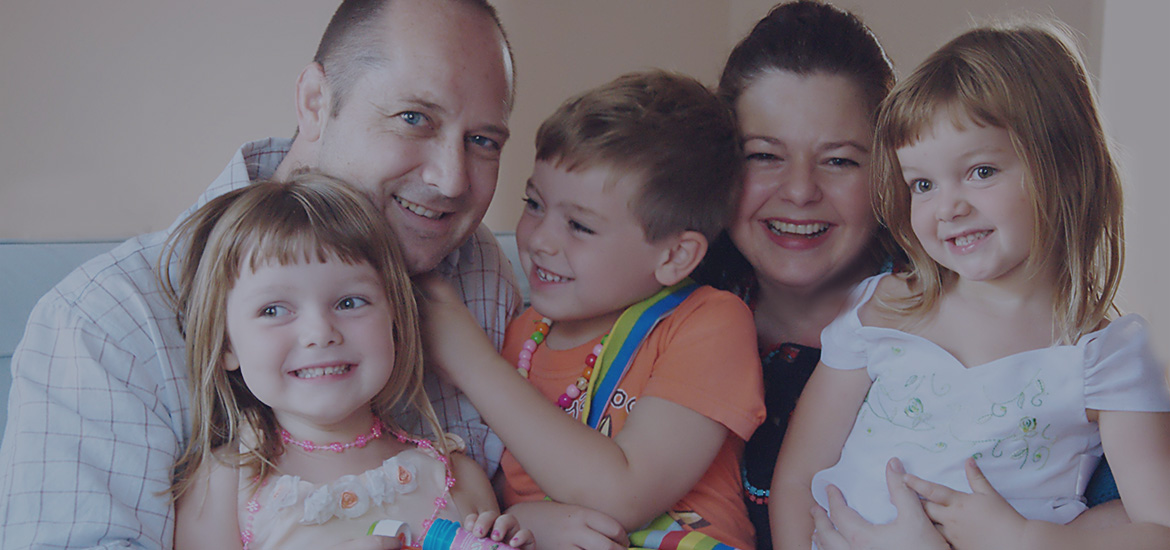
*
  
*
*
* *
Login 
  Friday, November 21, 2008About Cardiac Inherited Heart Disease 
 About Cardiac Inherited Heart Disease Minimize

         Most of these familial conditions have autosomal dominant transmission, with variable clinical expression.

         Thus half of the family members, on average, will carry the defective gene, but many will not have overt  features of the disease.

         Long QT / Brugada Syndrome

Long QT Syndrome (LQTS)

LQTS is characterised by abnormally prolonged ventricular repolarisation due to defects in cardiac ion channels. Prolongation of ventricular repolarisation is shown on the surface electrocardiogram by prolongation of the interval between the onset of the Q wave and end of the T wave. The condition predisposes affected people to tachyarrhythmias, which may lead to sudden loss of consciousness or sudden death, most commonly with exercise or stress, but also at times of rest. This is particularly true in infancy, where the cardiac sodium channel encoded by the gene SCN5A accounts for about half of infant deaths related to long QT syndrome [Arnestad 2004]; whereas it occurs in about 8% of the older population presenting with long QT syndrome [Splawski 2000].

LQTS is inherited as an autosomal dominant condition, with 10 candidate genes currently identified.  Predominantly, mutations in three genes encoding proteins of cardiac potassium and sodium ion channels have been identified: KVLQT1, HERG, and SCN5a which account for ~ 35%. 25% and 10% of cases respectively [Curran 1995; Wang 1995; 1996; Russell 1996; Splawski 2000; Ackerman 2004].  Approximately 60% of known families have identified mutations.  The frequency of these cardiac channelopathies may be greater than 1/2000 of the population [Priori AHA 2005].

In our cohort of 53 gene-positive probands from New Zealand, mutations within KVLQT1 (KCNQ1) predominate (57%), followed by HERG (28%) and then SCN5A (15%).  SCN5A mutations have only been identified from sudden death or “near miss” victims. Genetic diagnosis now plays a pivotal part of management and counselling of these families [Priori NEJM 2003]. Clinical diagnosis in family members may be uncertain in as many as 50%.  In families where a mutation has been identified, 30% of mutation carriers have a normal electrocardiogram.  They are, nevertheless, at risk for adverse cardiac events.  Administration of beta blocker medication can reduce the chance of sudden death by up to ten-fold [Moss 2000]. Other effective strategies include avoidance of triggers linked to the genotype (such as swimming in KVLQT1, and loud alarm clocks with HERG), avoidance of certain medications, and in a minority, defibrillator pacemakers or cervical sympathectomy.

Brugada Syndrome

Brugada syndrome was first described in 1992 as a form of idiopathic ventricular fibrillation associated with a persistent ST-segment elevation and a right branch block (RBBB) pattern on the electrocardiogram [Brugada and Brugada 1992].  These patients are susceptible to developing life-threatening ventricular fibrillation particularly during sleep [Chen et al 1998, Corrado 2001, Vatta 2002].  Although incidences are not well established, in South-East Asia, Brugada syndrome is the leading cause of death of young adults, exceeded only by deaths in vehicular accidents [Nademanee et al 1997]. 

The gene responsible for this disorder is SCN5A, the same gene that causes long QT type-3 and a cardiac conduction disease [Wang et al 1995; Tan et al 2001].  However, what determines the outcome of SCN5A mutations is the position of the mutation in the gene and the resulting differences in the electrophysiological abnormalities of expressed proteins.  We recently reported the first infant in whom Brugada syndrome (also known as SUNDS- sudden unexpected nocturnal death syndrome) has been linked unequivocally to a near- miss sudden infant death (Skinner 2005). As this infant’s resting ECG was completely normal, identification of the mutation has facilitated the finding of carrier status in the mother and sibling.

We have also recently described a 21 month old girl whose seizure-like presentation with fever was initially mistaken as a febrile convulsion- a relatively harmless childhood condition (Skinner 2007). In fact she had ventricular tachycardia, and she has Brugada syndrome. Fever is now recognised to be the commonest cause of collapse or blackout in children with Brugada syndrome (Probst 2007). 

  • Catecholaminergic Polymorphic Ventricular Tachycardia (CPVT)

CPVT is a rare arrhythmogenic disease characterised by exercise or stress induced ventricular arrhythmias, syncope or sudden death, usually in the paediatric age group, though it is not yet described in infancy [Tester 2004, Francis 2005]. Familial occurrence has been noted in at least a third of cases, and inheritance can be dominant or recessive, with high penetrance. Like LQTS, CPVT can be mistaken for epilepsy and be treated, inappropriately, with long-term anticonvulsants.  Moreover, similar to LQTS type 1, swimming with CPVT can trigger arrhythmogenic events, and is suspected as an important cause of unexpected drowning [Ackerman 1998, Moss 1999, Choi 2004].  The resting ECG is normal.  Exercise test, Holters or digital loop recorders may reveal polymorphic ventricular tachycardia during exercise (Coumel 2002), though this is not completely reliable.  The mainstay of treatment is beta blockade, which certainly reduces the episodes of VT on Holter (Coumel 2002). However on follow up of patients receiving beta blockade over 7 years, one group found a mortality of 10% [Leenhardt 1995], whilst another found 25% [Sumitomo 2003]. Missing a single dose of beta blocker can be fatal, and defibrillator pacemakers are being used more frequently.

Mutations within the cardiac Ryanodine gene (RyR2) have been identified in autosomal dominant pedigrees [Priori 2001, Laitinen 2001]. Three different isoforms have been identified, however, it is ryanodine receptor type 2 (RyR2) that is expressed in cardiac muscle. RyR1 is the skeletal muscle ryanodine receptor that is involved in malignant hyperthermia [Tiso 2001]. The receptors share a common molecular structure, whereby channels are formed as homotetramers composed of four RyR2 polypeptides. The Ca2+ channels regulate excitation-contraction coupling in cardiac muscle. Normally, the receptor is regulated by a range of accessory proteins resulting in normal Ca2+ homeostasis. The RyR2 is a large gene, made up of 105 exons, but to date mutations have only been identified within three distinct regions of the gene, covering 40 exons [tester 2004]. Point mutations within RyR2 account for CPVT in at least 38% of cases [Laitinen 2001]. The mechanisms by which mutations in the cardiac ryanodine receptor sometimes lead to catecholamine-mediated ventricular tachycardia and sometimes to ARVC are not clear. CASQ2- a calcium binding protein located in the sarcoplasmic reticulum – has also been implicated in a severe and uncommon autosomal recessive form of CPVT, [Postma 2002, and Lahat 2001].

In a recent screening study of DNA from 49 victims of sudden death with a negative post-mortem, with a mean age of 14 years, revealed that 7 (14%) had pathological mutations within RyR2 [Tester 2004].  The youngest child was aged 2 years; he died suddenly whilst watching TV. No such study has to date been performed on SIDS victims. Two such children (aged 11 and 12) presenting with recurrent syncope associated with exercise or emotional excitement have, in the last year, received the diagnosis of CPVT in Auckland. Both children had documented polymorphic VT recorded with an implanted digital loop recorder and both now have implanted cardioverter defibrillators.

Screening for asymptomatic gene carriers is hampered by the fact that the resting ECG is normal. Exercise testing and Holter monitoring can be used, but the interpretation of the result in this context is difficult, particularly if there is a negative result. Compared to LQTS “the lethality associated with CPVT seems greater and it may represent the ideal arrhythmogenic killer that is able to escape suspicion, detection, and apprehension by either a standard medico-legal autopsy or a careful evaluation of surviving first and second degree relatives” [Tester 2004]. It would thus be very desirable to be able to offer genetic testing when the post mortem from a sudden death is negative, to facilitate screening within such families.

An excellent review aimed at clinicians from the Italian group led by Sylvia Priori is available- click here.


  • Hypertrophic Cardiomyopathy (HCM)

Cardiomyopathy is defined as a disease of the myocardium associated with cardiac dysfunction [Maron 2002]. Hypertrophic cardiomyopthy (HCM), is the most common cause of sudden cardiac death during vigorous physical activity, particularly in young athletes aged ≤39 years.1-3  This has received considerable attention in the lay and medical press with the unexpected sudden death of high profile athletes [Maron 1996, Varnava 2001, Firoozi 2002, Behr 2002]. Histological features of myofibril disarray may present in the absence of ventricular hypertrophy,11  and though myopathic changes on skeletal muscle biopsy have been reported in some individuals with HCM their relationship to the mutations are unclear [Doolan 2004].

Hypertrophic cardiomyopathy is a disorder affecting myocytes, and it has a frequency of ~ 1/500 in the general population [Maron 2002].  It is generally characterised by myocardial hypertrophy not attributable to a known secondary cause such as systemic hypertension or valvular aortic stenosis. Using two dimensional echocardiography, at least 50% of cases are familial though this may well be an underestimate due to de novo mutations and genetic transmission which has not been clinically appreciated.  The pattern of inheritance is generally autosomal-dominant with variable penetrance. Also, individuals within kindred may have similar echocardiographic features and different symptoms, or varying degrees of hypertrophy and haemodynamic obstruction with the same mutation.

Given the variety of symptoms leading to clinical presentation, providing prognostic advice to individuals and families, in the absence of a genetic diagnosis, is not straightforward. The risk of sudden cardiac death correlates with the degree of myocardial hypertrophy, and several other features as well as the degree of left ventricular outflow obstruction [Maron 2002].

The identification of the molecular genetic bases for this disorder started with identification of linkage association of chromosomal locus 14q1 to the clinical expression of HCM in a French-Canadian family, who were later shown to be a point mutation in the β-MHC gene [Fannanapizir 1994]. Subsequently >100 mutations have been found in this gene [Maron 2002].  Linkage analysis, however, suggested that familial HCM is a genetically heterogeneous disorder and no abnormalities in the β-MHC gene were found in seven affected French families. Subsequently at least 8 other sarcomeric proteins, particularly myosin binding protein C and troponin T, β-tropomyosin have been confirmed as having mutations in affected individuals from families with HCM. Mutations in the genes encoding β-MHC gene and myosin binding protein C gene each account for ~25% of all FHC cases. Additionally, the pathogenic mechanism that links various mutations in different genes to a clinically indistinguishable phenotype has yet to be elucidated.

Further information and lay summaries are available at the Cardiomyopathy Association of Australia.

Arrythmogenic Right Ventricluar Cardiomyopathy\ Dysplasia (ARVC)

Arrhythmogenic Right Ventricular Cardiomyopathy (ARVC) is characterised by fatty infiltration of the right ventricle, fibrosis and ultimately thinning of the ventricular wall with chamber dilatation.  Arrhythmogenic right ventricular cardiomyopathy (ARVC) has been reported to account for 17% of sudden death in the young in the USA (Towbin 2001). ARVC has been increasingly recognized as a cause of both ventricular arrhythmias, and of premature sudden cardiac death.  Since the clinical manifestations are heterogeneous diagnostic criteria have been developed, and the classic features are often not initially manifest. Also, left ventricular involvement can occur. 

It was first recognized that ARVC had a genetic basis, in the early 1980's when Marcus et al. 1982 described 2 families, each of which had several members affected. In the general population, the prevalence of ARVC has been reported to be 1/5000, but this is likely to be a significant underestimate, because diagnosis has been difficult (see discussion below), prior to the availability of magnetic resonance imaging (MRI). ARVC is a genetically heterogeneous disorder, though inheritance is usually autosomal dominant with variable penetrance. There are candidate genes at least 7 chromosomal loci. Two candidate genes have recently been identified 1) plakoglobin (JUP) gene, which has been reportedly associated with Naxos disease [McKoy 2000, Tiso 2001] and 2) and mutations of the Ryanodine receptor, RyR2 (see below). RyR2 interacts with four-12KD EF-hand calcium binding proteins to control release of calcium from the sarcoplasmic reticulum to the cytosol, and thus ARVC can be considered an inherited channelopathy.

  • Dilated Cardiomyopathy (DCM)

Dilated Cardiomyopathy (DCM) has a genetic basis in a proportion (~25%) of cases with mutations found in 10 genes encoding cytoskeleton proteins leading to dilatation of the left ventricle predominantly (Franz 2001). Echocardiography usually shows a dilated poorly contractile left ventricle, with accompanying dilatation of the right ventricle in some cases. 

DCM may show evidence on endocardial biopsy of myocarditis such as lymphocyte infiltration which may include killer cell attack on myocytes.  However, these biopsy features are non specific, as are other histological findings such as myofibril loss, variable interstitial fibrosis and hypertrophy of the remaining myocytes. In Western communities, the most common cause of DCM is due to coronary heart disease, however ~25% of cases are familial and the patients often develop symptoms at a much younger age than patients with coronary heart disease.  Several patterns of inheritance of familial DCM have been reported including autosomal dominant, autosomal recessive and X chromosome-linked.  X chromosome-linked DCM is typically characterised by onset of heart failure in affected males in their mid-teens to early twenties.  Affected individuals show a progressive deterioration to death within a few years in the absence of cardiac transplantation.  Female carriers may be asymptomatic but some develop impairment of left ventricular function in their middle age.

 In 1993, discovery of the dystrophin gene to linked to X-linked DCM [Muntoni 1993], as well as the identification of a mutation in the promoter region of the same gene was reported.  Clinical skeletal muscle weakness has not been observed in X-linked DCM patients with dystrophin gene mutations.  This observation may be due to the fact that there are at least four tissue-specific dystrophin promoters, and a mutation has been identified in the muscle promoter of the dystrophin gene by Muntoni et al.  Other mutations have been reported in other cytoskeletal proteins as well as dystrophin, which are the primary biochemical defect in Duchenne (DMD) and Becker (BMD) muscular dystrophies, which are disorders that present with skeletal muscle weakness in the first or second decades of life, respectfully, and occur in 1/4000 of the population, called dystrophinopathies.


 Print   


Copyright 2007 by Detec Consulting
*
Detec Ltd   Terms Of Use   Privacy Statement